The FMIAC is housed in MRB. It consists of equipment for obtaining digitized images of gels, blots, and microtiter plates for radioactivity, chemiluminescence and chemifluorescence in the Macroscopic Imaging Facility and for various light/fluorescence microscopy applications in the Microscopic Imaging facility.
Macroscopic Imaging Facilities
 Macroscopic Imaging Facility Supervisor: Dr. Santiago Di Pietro, Associate Professor
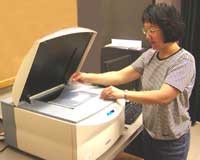
The macroscopic imaging equipment in room 210 MRB includes a Typhoon RGB scanner for radioisotopic, chemiluminescence and fluorescence imaging. Laser lines of 473, 532, and 635 are available and large format phosphorimager screens allow detection of radioactivity over a 105 dynamic range. An ImageQuant LAS 500 detection system for chemiluminescence, fluorescence and colorometric measurements from microtiter format plates is available in the same room. Across the hall in room 214 MRB is a LiCor Odyssey CLx near IR Scanner for dual color Western blot analysis. This equipment is not reserved but open to users on a first-come first-served basis. Users from outside the department need to be trained and have an account on file for hourly charges incurred.
Fees For Macroscopic Imaging Equipment: Odyssey costs are billed at $235/month divided by hours of use, averaged over 6 months. Typical hourly costs are between $25-30. Typhoon costs are billed at $450/month divided by hours of use, averaged over two month periods. Typical hourly costs range from $30-70.
Microscopic Imaging Facilities
Microscope Imaging Facility Supervisors: Zeiss LSM 900 Microscope, Olympus IX83 Spinning DisK Microscope and Keyence All-In-One Fluorescence Microscope: Alisa E. Shaw, Senior Research Associate, Tel. 1-5531; Nikon TIRF Microscope: Keith DeLuca, Research Associate, Tel: 1-5149; Nikon Diaphot: Laurie S. Minamide, Senior Research Associate, Tel: 1-5531.
The Microscope Imaging Center is housed in room 224 MRB. The fluorescence microscopes in this center are part of the CSU Microscope Imaging Network.
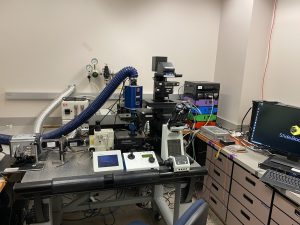
Zeiss LSM 900 Confocal Microscope with Airyscan 2
Fully automated inverted laser scanning confocal microscope (Axio Observer 7) with superresolution capability and Definite Focus 3. The system has four diode lasers (405, 488, 561 and 640 nm) and differential interference contrast (DIC) optics with 10X, 20X, 40X, and 63X objectives. A motorized XYZ stage and environmental chamber with
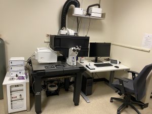
Zeiss LSM 900 Microscope
temperature, CO2/air and humidity control supports culture dishes, multi-well plates or slides. The system has two MA photomultipliers and one GaAsP Airyscan 2 photomultiplier detectors. The Airyscan 2 detector provides a combination of gentle superresolution imaging with high sensitivity. Airyscan 2 is an area detector consisting of 32 concentrically arranged detection elements. The confocal pinhole remains open, thus collecting more photons and resulting in much greater light efficiency. The multiplex mode allows for parallel pixel acquisition for faster frame rates, which facilitates observation of highly dynamic processes in living specimens. The system allows for photobleaching, photoactivation or photoconversion of user defined regions of interest while imaging, which is ideal for experiments such as FRAP. A Colibri 7 LED light source, filter sets and Axiocam 506 camera facilitate specimen visualization for quick identification of regions of interest before laser scanning confocal imaging. The microscope head is surrounded by a stabilization chamber (XLmulti S2 DARK Premium) that controls temperature and precludes room light from affecting the specimen or image acquisition. A full Zen Blue System Software package controls the microscope and contains a variety of analysis tools. The system is placed on a vibration isolation air table and connected to RAID storage.
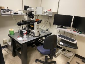
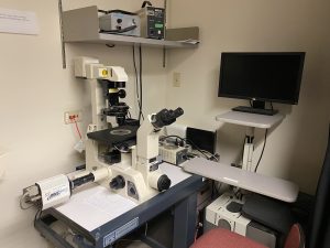
Keyence Microscope
Keyence All-in-One Fluorescence Microscope BZ-X710. The fully enclosed stage chamber of this Keyence microscope allows all imaging to be done under normal room lighting conditions. The microscope is fully under computer mouse control for X,Y,Z stage, filter cube rotator and turret objective switching device. It automatically maps out viewing area for slides, dishes and titerplates. Filter cubes (4) are easily switched in and out during observations without the need to turn off the light. Standard configuration has DAPI, GFP, mRFP and an open position for brightfield but cubes are available for Cy5/iRFP (640 line on laser systems) and NeuO, a neuronal viability stain that is excited at 488 and emits at >620 nm. In brightfield mode, oblique illumination is used to achieve near DIC images. A stage incubator for temperature and humidity control is also available. Standard objectives (all Nikon) include 2x, 10x, 20x, 40x, 40x long working distance (LWD), and 100x oil. Although not a confocal system it performs image stacks at near confocal resolution with a minimum of 0.1 µm steps. It will obtain stacks through 500 µm thick slices. The unit has a high sensitivity cooled monochrome CCD that is converted to a 3CCD color camera for staining images at the click of a button. A large motorized stage, high-speed autofocus, low photobleaching mode and real-time image overlays are features of the core microscope. Advanced imaging functions that are all available include image stitching in either an autofocus mode for each image (slow) or full focus wide-area (about 30 sec to scan a single plane human hippocampal slice in brightfield and display the montage), navigation (maintains image of montage on side panel and lets you see on the montage where you are located for higher magnification imaging), optical sectioning, and time-lapse. A structured illumination module use projection of an optical grid to achieve super-resolution fluorescence images.
A Peltier cooled stage adapter is available for use on this microscope. It will allow imaging of cells in 35 mm glass bottom dishes (without the double thick plastic walls at the base). This device will cool cells in medium at approximately -20o C/min to below 0o C, although the precise rates of cooling need to be determined for the volume of medium. Continuous fluorescence imaging (time lapse/multi point) can take place while cooling. Training is required for this application since no specific stage adapter is yet available.
Analysis software is included on the system and an off-line package is also available on a second computer in the facility. Applications include hybrid cell counts (can use dual fluorescence or brightfield/fluorescence to automatically quantify the % of cells expressing tags along with color extraction (for stained sections) and masking functions for selection of regions of interest. A macro cell count application allows parameters set for one acquisition to be applied to multiple samples, especially useful for batch processing and high throughput analysis. A measurement module allows point and click 2D measurements for length, distance, area, etc. and a 3D module allows visualization fluorescence image stacks with rotation, zoom in and out functions, sectional views, XYZ slicing and maximum projections.
Nikon TIRF Microscope
Nikon Total Internal Reflection Fluorescence (TIRF) Inverted Microscope (Eclipse Ti) has perfect focus control, a four laser line launch (405, 488, 561 and 640 nm) and an Intensilight Hg illumination source. A motorized XY Piezo Z stage and stage-top environmental chamber with temperature, CO2/air, humidity capability, will support culture dishes, multi-well plates or slides. Equipped with 20X, 40X, 60X DIC objectives and a 100X TIRF objective. Microscope has an Andor Clara camera for wide field imaging and an Andor iXon3 EMCCD camera for TIRF imaging. Microscope has built-in N-STORM super-resolution imaging capability and is networked to a RAID array for multiple terabyte storage. Nikon elements software controls all aspects of acquisition and analysis with a deconvolution software package included.
Olympus IX83 Spinning Disk Confocal Microscope
The Olympus IX83 Inverted Spinning Disk Confocal (SDC) Microscope was recently upgraded from an IX81 microscope frame to include a Zero Drift Compensation (ZDC) autofocusing unit. The microscope has a custom-built stage top environmental control chamber with CO2 control for live cell work, an X,Y, piezo Z stage for rapid image stack acquisition across many predetermined fields (4 D imaging), a CSU 22 head with quad dichroic and additional emission filter wheel to eliminate spectral crossover, four high power diode lasers (405 nm, 488 nm, 561 nm and 647 nm) with rapid (microsecond) switcher and a phasor holographic photobleaching/photoactivation/photoconversion system for intracellular molecular dynamic measurements. Phasor can also be used for ion uncaging and other applications. System has differential interference contrast (DIC) optics with 10, 20, 40, 60 and 100X objectives, built in correction for spherical aberration for all objectives, and a wide field Xenon light source. A cascade II EMCCD camera (confocal imaging) and a Photometrics HQ camera (wide field imaging) are both integrated for image capture using Slidebook software. Laser selection and power are software controlled. System is connected to RAID terabyte storage.
Nikon Diaphot Epifluorescence Microscopes
Nikon Diaphot inverted epifluorescence microscopes (two) with video cameras for digital imaging are also available for routine fluorescence imaging on fixed samples. Filter cubes for all common fluors (DAPI, Fluorescein, Rhodamine, Texas Red, Cy 3, Cy5) are available and are interchangeable between microscopes. One microscope has DIC optics, an optional phase oil-immersion condenser, and a stage incubator and filter wheel for live cell studies (ratio imaging included) and one has a color camera for imaging stained sections. A specialized light source for darkfield illumination of slides is available and is especially useful for counting silver grains on emulsion treated samples. All microscopes utilize Metamorph software for image capture and analysis.
Data Storage
Users must bring their own drives for downloading image files as soon as the experiments are complete. Storage capacity for a longer time may be purchased on the Terabyte drive system.
Fees for Spinning Disk and TIRF Microscope Equipment: Training: Cost for training is $250, which includes 5 hours of microscope access after training. Short-term use: $20 per hour flat rate. Long-term use: New users pay $20 per hour up to a maximum of $400 for the first month, $300 for the second month and $210 per month thereafter. The increased rates for the first two months reflect the additional supervisory time and maintenance/service required for new users. Costs decline as users become proficient in application of the many different items of ancillary equipment and program applications associated with the microscopes. Fees for the Diaphot: $3.50 per hour.
Schedule Time: Confocal; Keyence
Access data on NAS1: Web Mac OS X On Windows, map network drive to \\fmic-nas1.bmb.colostate.edu\home
E-mail Userlist: Confocal
